
You notice a rounded chest shape in the mirror, or a tender spot beneath one nipple when your shirt brushes against it. Maybe you've already tried changing your training or losing weight, but a firmer area remains. The practical question is how to tell if you have gynecomastia, rather than ordinary chest fat, and when that difference deserves a medical evaluation.
Gynecomastia is common, but common doesn't mean every new lump should be dismissed. The texture, location, timing, symmetry, tenderness, and associated symptoms all matter. A self-check can help you describe what you're feeling, but only a clinical examination can reliably distinguish normal variation from glandular tissue, medication effects, hormonal disease, or a less common serious cause.
Table of Contents
- What Gynecomastia Actually Is and Who It Affects
- How to Perform a Simple Self-Check at Home
- True Gynecomastia vs Pseudogynecomastia
- Red Flags That Mean You Should See a Doctor Sooner
- What to Expect During a Medical Evaluation
- Treatment Options Including Awake Male Breast Reduction
- Your Next Steps and Key Takeaways
What Gynecomastia Actually Is and Who It Affects
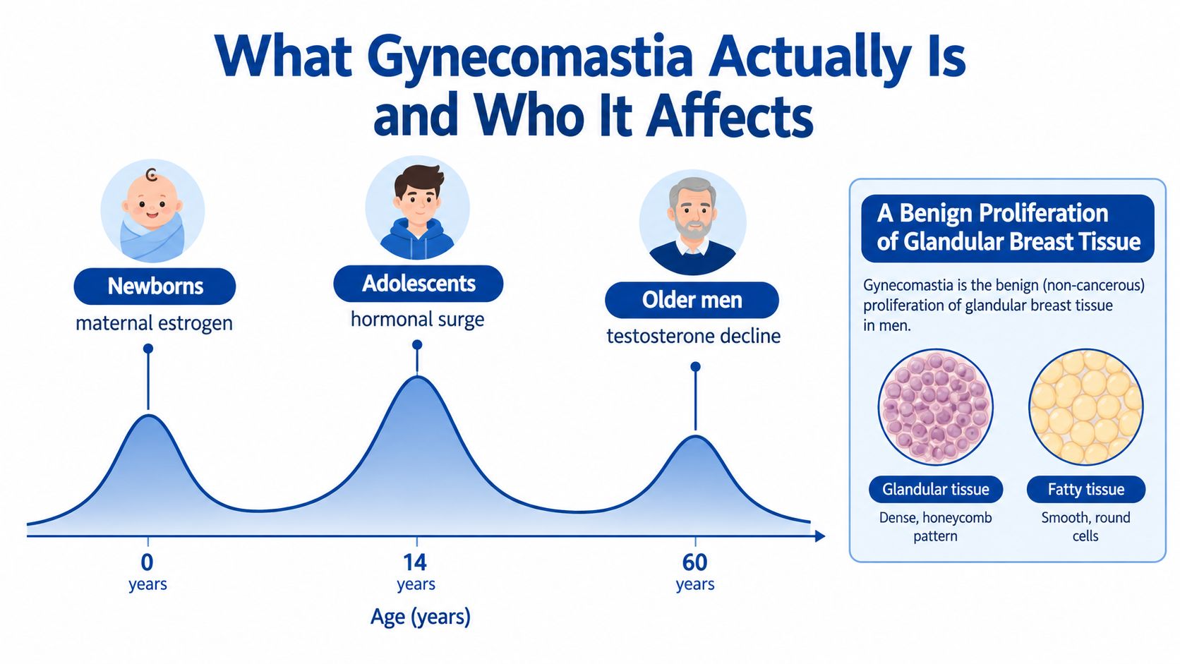
A newborn may develop a small breast bud after exposure to maternal hormones. A teenager may notice a tender disc beneath one nipple during puberty. An older man may develop gradual enlargement as androgen activity and hormone metabolism change. These are the three age-related periods in which gynecomastia, benign growth of glandular breast tissue in males, appears most often.
The tissue is different from fat spread across the chest. It forms beneath the areola and may feel firm, rubbery, or sensitive. Reported prevalence is 60–90% in neonates, 50–60% in adolescents, and up to 70% in men aged 50–69 years, according to a clinical review of gynecomastia.
Pubertal enlargement commonly settles as maturation continues. By age 17, only about 10% of boys have persistent gynecomastia, so a teenage lump can be a normal hormonal change when its timing, texture, and symptoms are reassuring. Persistence does not automatically mean disease, but it warrants assessment if the pattern is unusual.
In adulthood, a relative shift in the estrogen-to-androgen balance can stimulate ductal and supporting tissue beneath the areola. At least 30% of males are affected at some point in life. New or progressively enlarging tissue, marked pain, one-sided development, medication exposure, or other atypical findings change how the evaluation should proceed.
Spironolactone, finasteride, and some psychiatric medicines can contribute. Anabolic steroids, marijuana, liver or kidney disease, hypogonadism, and other endocrine conditions may also alter the picture. Body composition affects the examination: substantial chest fat can hide a smaller glandular disc, while leaner patients may notice its firm border immediately.
If enlargement remains after a likely trigger is addressed, or it affects confidence, a male breast reduction and gynecomastia consultation can clarify whether the chest contains gland, fat, or both, and whether surgery is appropriate.
How to Perform a Simple Self-Check at Home
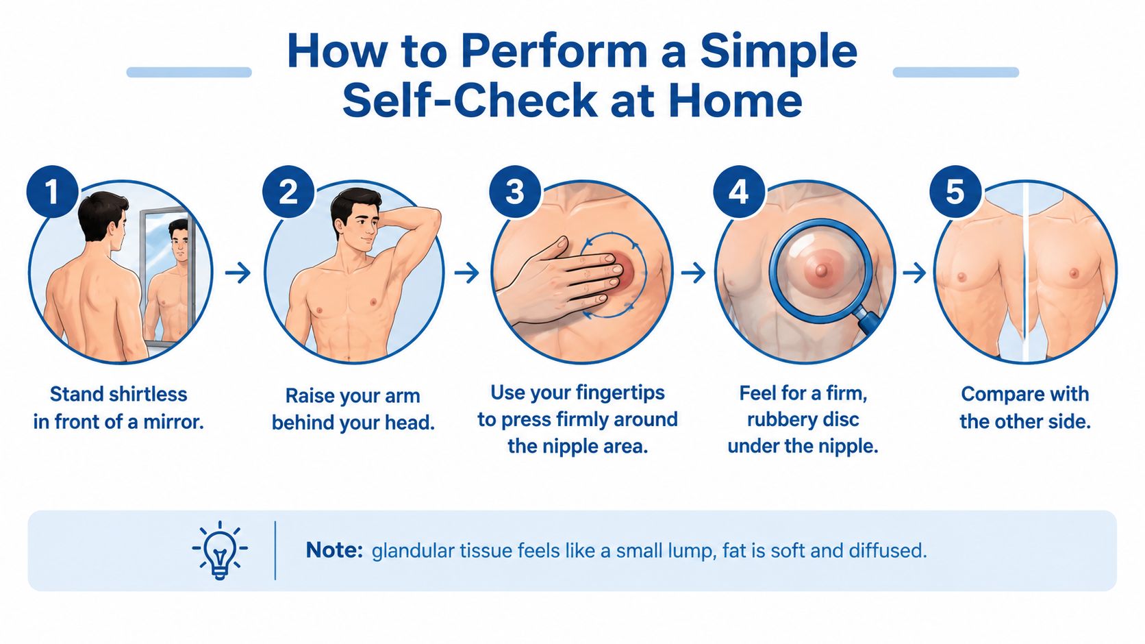
Start shirtless in front of a mirror with your arms relaxed. Look for a rounded mound, puffy areola, nipple displacement, skin changes, or a clear difference between sides. Then raise your arms overhead. This position stretches the skin and can make a central bulge, indentation, or asymmetrical contour easier to see.
Use the pads of your index and middle fingers rather than pinching hard with your nails. Place them flat against the edge of the areola and gently press inward toward the chest wall while moving in small circles. Work from the outer tissue toward the nipple, then repeat from several directions. The aim isn't to squeeze aggressively. Excess pressure can create soreness and make normal tissue feel abnormal.
What your fingers may find
True gynecomastia usually feels like a firm, rubbery, discrete disc or button directly under the nipple. It may be mobile against the deeper chest wall, and early tissue can be sensitive or tender when touched. The border often feels different from the surrounding tissue, which is the feature that separates it from diffuse fat.
Pseudogynecomastia feels more like the rest of the body's fat. It's generally soft, doughy, compressible, and spread across the chest without a defined central plate. A higher body-fat level can make the examination harder because fat may surround a smaller glandular disc. In that situation, you may feel both a soft layer and a firmer area beneath the areola.
Practical rule: Compare both sides, but don't assume they must match. Gynecomastia can be one-sided or uneven, especially early in its development.
Repeat the examination lying down if you can do so comfortably, because a structured clinical breast examination is performed with the patient supine and palpation directed toward the nipple-areolar complex clinical examination guidance. Make a private note of the location, approximate extent, tenderness, and any nipple or skin change. Consistent photos under similar lighting can help you notice progression, but they can't diagnose the tissue.
True Gynecomastia vs Pseudogynecomastia
Consider two typical presentations. A lean gym-goer notices a mildly sore, rubbery area beneath one nipple shortly after starting finasteride. The timing, focal disc, and tenderness make true gynecomastia more plausible, although the medication shouldn't be stopped without guidance from the prescribing clinician.
A different man notices gradual fullness across both sides of the chest. He has a higher body-fat level, feels no distinct lump, and sees the chest become less prominent as his overall weight decreases. That pattern points more toward pseudogynecomastia, which is fat rather than glandular proliferation.
Mixed cases are common. A man can have a glandular disc beneath each areola and a broad layer of fat over the chest. That combination matters because the procedures that address each tissue type aren't interchangeable. Liposuction removes fat, while direct excision removes firm glandular tissue. Removing fat alone may leave a central disc behind, while excision alone may leave a soft fatty halo.
| Feature | True Gynecomastia | Pseudogynecomastia |
|---|---|---|
| Tissue | Firm glandular tissue beneath the areola | Diffuse fatty tissue |
| Feel | Rubber-like disc or button with a defined edge | Soft, doughy, and evenly compressible |
| Location | Usually centered under the nipple | Spread across the chest |
| Tenderness | Can be sore, particularly when recent | Usually not tender unless skin irritation or trauma is present |
| Response to weight loss | The glandular component often remains | Fatty fullness may decrease with overall fat loss |
| Treatment implication | May require direct excision, sometimes with liposuction | Lifestyle changes or liposuction may address the fat component |
A self-check can suggest a pattern, but it can't rule out malignancy or another abnormal process. Any hard, fixed, eccentric, rapidly enlarging, or skin-changing mass needs medical assessment regardless of body composition.
Red Flags That Mean You Should See a Doctor Sooner
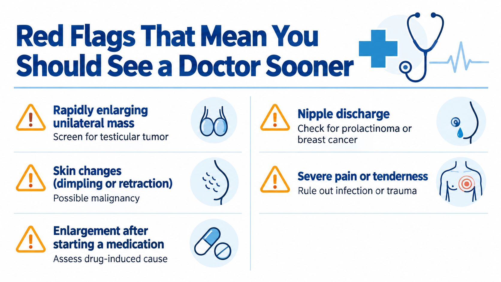
A new lump that grows quickly, appears mainly on one side, or feels unlike the usual soft fullness deserves a clinical assessment. This pattern can reflect a hormonal source outside the breast, including a testicular problem, or less commonly a male breast tumor. The workup may include a testicular examination, hormone testing, and breast imaging, depending on the history and examination.
A hard, fixed, non-tender mass away from the nipple is more concerning than a soft, centered disc beneath the areola. Skin dimpling, nipple inversion, ulceration, enlarged lymph nodes, or spontaneous discharge, especially bloody discharge, also change the evaluation. These findings do not prove cancer, but they should not be watched casually.
Findings that alter the clinical pathway
- Rapid growth or marked asymmetry: A sudden change can prompt targeted breast imaging and endocrine testing, particularly when the tissue does not feel like a typical subareolar gland.
- Testicular swelling or a palpable testicular change: Palpation can miss some testicular tumors. Testicular ultrasound is appropriate when clinical uncertainty or abnormal hormone findings exist evaluation of gynecomastia and testicular causes.
- Nipple discharge or skin retraction: These features require assessment for breast pathology rather than reassurance based only on age.
- Severe pain, recent trauma, or bruising: Injury can produce an inflammatory fat lesion that resembles gynecomastia during examination.
- Jaundice, unexplained weight loss, or known liver or kidney disease: These clues may indicate altered hormone metabolism or another illness contributing to breast enlargement.
- Persistence in an adolescent: Enlargement that continues beyond the usual observation window of 12 to 18 months should be reviewed instead of repeatedly dismissed as normal puberty adolescent gynecomastia review.
- New enlargement later in life: Onset after age 60 calls for a careful history and examination. Medication effects, endocrine disease, systemic illness, and malignancy all become relevant possibilities.
Do not keep pressing the area in hopes of reaching a definite answer when a red flag is present. Arrange a focused clinical evaluation, particularly if the change is new, one-sided, painful, or associated with testicular or systemic symptoms.
What to Expect During a Medical Evaluation
A medical visit begins with questions that clarify the pattern, timing, and possible cause of the enlargement. Your clinician will ask when it started, whether it has progressed, whether one or both sides are affected, and whether the area is painful. Bring a complete list of prescription medicines, supplements, performance-enhancing substances, and recreational drugs. That history can reveal a trigger that the chest appearance cannot.
The discussion may also include alcohol use, thyroid symptoms, liver or kidney disease, changes in sexual function, testicular symptoms, and family history of breast cancer. These questions are routine, not judgmental. The answers help determine whether observation, blood tests, imaging, medication review, or specialist referral makes sense.
What the physical examination involves
The clinician first inspects your chest while you are sitting or standing. They assess symmetry, nipple position, skin changes, and the overall contour. You may then lie back with an arm behind your head while the examiner palpates from the outer chest toward the areola.
The examination distinguishes a firm, rubbery, concentric disc beneath the nipple from diffuse, softer fat. Age and body composition affect how clear that distinction feels. In a lean adolescent, glandular tissue may be easy to identify. In a heavier adult, fat can surround the gland and make the borders less obvious. The examiner also checks whether a lump is hard, fixed, eccentric to the nipple, or associated with skin or nipple changes.
The breast and genital examinations should be considered together. Testicular palpation can miss a lesion, so scrotal ultrasound may be appropriate when the examination is uncertain or hormone results suggest a testicular source. Clinicians may record the amount of gland, fat, and loose skin with a recognized grading system, which helps match treatment to the actual anatomy.
| Test | What it screens for |
|---|---|
| Testosterone, estradiol, LH, and FSH | A disrupted sex-hormone pattern or impaired testicular function |
| Prolactin | A hormonal cause associated with abnormal prolactin activity |
| hCG | A possible hormone-producing tumor source |
| TSH | Thyroid dysfunction that may influence breast enlargement |
| Liver function testing | Liver disease and altered hormone metabolism |
| Kidney function testing | Kidney disease and systemic contributors |
| Breast ultrasound or mammography | Atypical, hard, fixed, unilateral, or uncertain breast tissue |
| Scrotal ultrasound | A testicular lesion when examination or hormone findings raise concern |
Imaging is not automatic for a typical pubertal or medication-related presentation. The final plan should state whether the pattern appears physiologic, pharmacologic, idiopathic, or linked to another condition, and when reassessment is needed.
Treatment Options Including Awake Male Breast Reduction
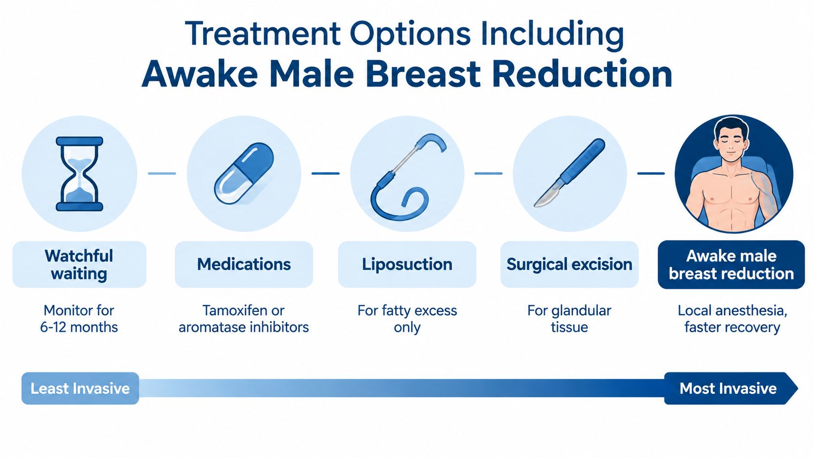
Treatment starts with the cause and the tissue type, not with a procedure chosen from a photograph. Early pubertal cases are often observed because many settle as hormones mature. If a medication, supplement, anabolic agent, or systemic illness is contributing, the safer first move is review and treatment of that trigger under medical supervision.
Medication can have a role in selected, recent cases while the tissue is biologically active. Selective estrogen receptor modulators and aromatase inhibitors have limited evidence and are less useful once long-standing tissue has become dense and fibrous. They aren't a substitute for evaluating a suspicious mass or correcting an underlying endocrine disorder.
Matching the procedure to the tissue
When the glandular component has stabilized and remains painful, visible, or psychologically distressing, surgery offers the most direct way to remove it. Liposuction alone works for fatty excess, not for a firm glandular disc. Direct excision is used for the gland, and many men need both techniques because the chest contains gland plus surrounding fat.
Awake male breast reduction can be a logical option for appropriately selected patients. The surgeon uses tumescent local anesthesia, with oral relaxation when appropriate, then removes the gland through a small periareolar incision. Liposuction can refine the fatty perimeter and help blend the treated area into the chest.
Potential advantages include avoiding general anesthesia and an intravenous line, a shorter recovery pathway, and the ability to assess contour with the patient positioned during treatment. The trade-off is that awake surgery isn't right for every anatomy or every patient. Large skin excess, significant drooping, extensive tissue, or a need for skin tightening may be better managed with general anesthesia and a more involved operation.
The best technique is determined by what I can feel and see, not by the label alone. A patient with a small gland and good skin tone may need a very different plan from a patient with substantial fat and loose skin.
Ideal candidates for an awake approach generally have gland-predominant enlargement, reasonably elastic skin, a healthy body composition, and no significant ptosis. A consultation at Ideal Face & Body's gynecomastia treatment options page can help explain whether gland excision, liposuction, a combination, or a different surgical plan fits the examination.
Your Next Steps and Key Takeaways
Use this week to collect useful information rather than repeatedly checking the same spot.
- Record the timeline. Note when you first saw the change, whether it is growing, and whether one side is different.
- Perform one careful self-check. Look for a firm, centered disc beneath the nipple and compare both sides without squeezing aggressively.
- Review exposures. Write down prescriptions, supplements, anabolic substances, and recreational drugs, then discuss them with a clinician before making changes.
- Screen for red flags. Rapid enlargement, a hard fixed mass, skin dimpling, nipple inversion, discharge, testicular changes, or systemic symptoms should move the appointment forward.
- Arrange an evaluation when it persists. Enlargement lasting beyond a year, ongoing pain, or uncertainty about the tissue is a good reason to schedule a primary care or surgical consultation.
The central distinction remains simple. True glandular gynecomastia generally won't disappear through diet or exercise alone, although reducing body fat can improve the fatty component around it. Pseudogynecomastia, by contrast, often becomes less prominent as overall body fat decreases.
Persistent glandular tissue doesn't automatically mean surgery is required, but it does make a discussion about realistic options worthwhile. If an operation becomes appropriate, ask how the surgeon will address the gland, surrounding fat, skin quality, anesthesia plan, symmetry, and recovery. Information about gynecomastia surgery recovery can help you prepare more specific questions.
Accurate self-identification is only the starting point. A hands-on examination and targeted workup turn suspicion into a diagnosis, then into a plan that fits your age, symptoms, tissue composition, and goals.
Ideal Face & Body offers private consultations for men who want to determine whether chest fullness is glandular tissue, fat, or both. Visit Ideal Face & Body to discuss an awake male breast reduction evaluation and the treatment approach that matches your examination.