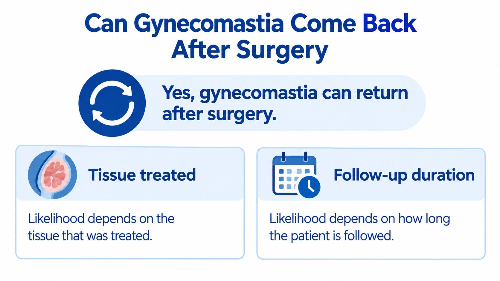
Yes, gynecomastia can come back after surgery.
Returning chest fullness can come from three different sources: true glandular regrowth, tissue left behind at the first operation, or new fat in the chest. Long-term follow-up has found very different recurrence patterns depending on whether the treated chest was mostly firm gland or mostly fat.
That difference matters because each cause has its own fix. Gland works like a root, while fat behaves more like a refillable cushion. If you label every returning fullness as “failed surgery,” you can chase the wrong treatment.
Table of Contents
- Can Gynecomastia Come Back After Surgery
- Residual Tissue and Active Causes
- Prevention Steps That Lower Your Risk
- Warning Signs and What Each One Means
- Revision Options and Awake Approaches
- Confirm the Cause Before You Book Revision
Can Gynecomastia Come Back After Surgery
Recurrence risk is written into the tissue, not the calendar. A chest built mostly of firm gland ages differently after surgery than a chest built mostly of fat, so a single recurrence percentage quoted without that context can send you chasing the wrong explanation.
What the long-term evidence shows
A study published in 2017 followed surgically treated men for well over a decade and found that recurrences clustered heavily in the fatty-type group, while changes in body-mass index stayed similar across groups. That pattern carries a useful lesson. In a lipomatous chest, returning fullness may be renewed fat rather than a regrown gland. Removed gland stays removed, but fat can accumulate again and redraw a similar outline, and each explanation calls for a different correction.
Recurrence by tissue type at long-term follow-up
| Tissue Type | Mean Follow-Up | Recurrence Rate |
|---|---|---|
| Glandular gynecomastia | 13.8 years | 12.5% (1 of 8) |
| Lipomatous gynecomastia | 13.8 years | 62.5% (5 of 8) |
Source: the 2017 long-term follow-up study, which reported the difference between tissue types as statistically significant.
Recurrence can also appear far sooner than a decade. A case report indexed by PubMed described unilateral gynecomastia recurring 3 years after subcutaneous mastectomy, liposuction, and tamoxifen, requiring a second operation, as documented in the PubMed case report record. That same case was later summarized in a 2024 review in the Journal of Men's Health, which reported recurrent gynecomastia 3 years after liposuction, mastectomy, and tamoxifen, followed by repeat surgery.
A clean first year is reassuring, but it doesn't prove recurrence is impossible over a longer horizon.
One detail shapes everything above: your original diagnosis. Whether your chest was mainly glandular or mainly fatty is the single most useful fact for interpreting any change you notice now, and it's worth confirming in your surgical records before you draw conclusions from the mirror.
Residual Tissue and Active Causes
A pruned hedge looks finished, but roots left in the ground can send up new shoots months later. Surgery works in a similar way. It removes existing tissue, yet it does not switch off the hormonal signals that made the tissue grow, and any responsive gland left behind can still react to those signals.

Three different problems can produce returning fullness: true regrowth of remaining gland, tissue that was never fully removed, and renewed fat. The fat-regain pattern described earlier is the third of these. The two gland-related causes deserve a closer look here, because they arise at different times, for different reasons, and they call for different corrections.
When tissue was left behind
Residual tissue is the first explanation to rule out. Liposuction alone cannot remove firm fibroglandular tissue, so whatever stays behind can remain prominent or enlarge later. In a review of severe cases, the reviewed literature reported postsurgical recurrence rates as high as 35% in this context, according to the severe gynecomastia literature review.
That same analysis traced the problem to surgical balance. Removing too little fibroglandular tissue produces undercorrection, a problem of what remained. Taking too much of the tissue that supports the chest wall produces overcorrection, a problem of what was lost. Adequate surgery has to clear the firm gland while preserving enough contour-supporting fat for a natural shape, and the margin between the two errors can be narrow.
When the original cause is still active
Sometimes the operation was technically sound, but the stimulus behind the original growth never stopped. Incomplete excision can leave tissue that remains responsive to endogenous hormonal stimulation, and operating before puberty has fully finished can permit new growth, as noted in clinical guidance on active causes after gynecomastia surgery.
The mechanisms differ, but they share one feature: each keeps feeding responsive tissue after the operation. Hormonal shifts such as higher estrogen can restart growth, and the evidence supports that link. In a retrospective cohort of 79 surgical patients, 26 experienced relapse and 53 did not, and higher estrogen levels and greater glandular-tissue thickness were associated with increased recurrence, with recurrence more common after minimally invasive procedures, according to the retrospective cohort study of 79 patients. Certain prescription medicines, anabolic-androgenic steroids, hormone-boosting supplements, and untreated liver, kidney, testicular, or endocrine conditions can each supply a continuing stimulus. Sustained weight gain can act on both fronts at once, adding chest volume and raising estrogen activity through the new fat tissue.
That is why apparent recurrence may reflect ongoing disease activity rather than technical surgical failure. Studies also define recurrence differently, follow patients for different lengths, and cover different procedures, so no single percentage applies to every patient. An individualized assessment carries more weight than any headline number.
One question helps narrow the cause. If your chest never looked flat after the first operation, undercorrection is the likelier explanation. If it looked good and later enlarged, recurrence or renewed fat is likelier, and that simple memory gives your clinician a useful starting point.
Prevention Steps That Lower Your Risk
Prevention works best when you match the habit to the look-alike you are trying to avoid: gland returning, tissue left behind, or fat returning. Each step removes one reason the chest can change shape.

- Keep your weight inside a narrow, personal range. Choose a range you can maintain, then check on a steady schedule and after travel, illness, or training breaks. A slow drift is easier to correct early than after clothes and photos make it obvious.
- Build a dated medicine and supplement file. Save labels, start dates, dose changes, and the prescriber for each item, then review the file whenever treatment changes. Medication decisions stay between you and the prescriber.
- Keep any hormone plan supervised and disclosed. If steroids, boosters, or testosterone treatment are part of the picture, tell your surgeon and primary clinician before cycles restart and agree on monitoring. For questions to raise, gynecomastia advice from Cost Plus TRT can help you frame the discussion.
- Agree on testing triggers before you need them. Ask your clinician which change should prompt labs and which result would alter treatment, so testing answers a defined question rather than anxiety.
- Create a follow-up routine you can repeat anywhere. Use the same lighting, mirror distance, and front/side photos on a regular schedule, and store them beside your medicine file. Consistent comparisons beat memory.
- Discuss the intended tissue map before surgery or revision. Ask what will be removed, what fat will be left for contour, and how symmetry will be checked during the operation. Clear targets reduce preventable mismatch between expectation and result.
- Decide your escalation path while you feel well. Write down who to call first, which photos and records to send, and how quickly a new change should be reviewed. Prepared patients get clearer answers sooner.
Practical rule: Prevention lowers risk but cannot remove it.
Good habits shrink the space where regrowth, undercorrection, and fat regain can hide. They still leave room for examination when your body changes.
Warning Signs and What Each One Means
Each row below pairs a sign you might notice with its most likely cause and the sensible next action.
Symptoms, likely causes and next steps
| Sign | Most Likely Cause | What to Do Next |
|---|---|---|
| Gradual fullness over months after weight gain | Renewed fat in the chest | Track weight and ask about contour-focused correction |
| Firm, rubbery disc under the nipple | Residual or regrown glandular tissue | Clinical exam, with imaging if the clinician advises it |
| Tenderness, swelling or a firm lump | Hormonal, medication-related or other medical cause | Medical evaluation before any surgery |
| Enlargement on one side only | Asymmetric residual tissue or another cause | Prompt exam to rule out other conditions |
| Enlargement after starting or changing a medicine | Medication-associated breast growth | Review the medicine with the prescriber; do not stop it alone |
Not every postoperative concern is recurrence. A 2021 clinical review reported revision rates after gynecomastia surgery ranging from 0% to 14.1%, while recurrence estimates varied by procedure and follow-up duration, according to the 2021 clinical review of gynecomastia treatment. Relapse is only one explanation among several, alongside residual tissue, contour irregularity, and scar-related change.
Timing helps you interpret what you're feeling. Swelling and firmness are normal for several months after surgery, so a chest that's still settling isn't automatically a recurrence, a point covered in guidance on gynecomastia surgery recovery. Suspect recurrence instead when a previously flat chest enlarges again, or when a new firm disc or lump appears after healing seemed complete.
Don't assume renewed enlargement, tenderness, or a firm lump is ordinary fat gain. Get evaluated, since clinicians may review your medications, endocrine conditions, hormone levels, and original operative technique before considering revision. Write a dated timeline of when each change appeared and bring it to the appointment, because dates often reveal the trigger faster than memory does.
Revision Options and Awake Approaches
Match the revision to the diagnosis. Three different problems can produce a similar-looking chest: true glandular regrowth, residual tissue the first operation left behind, and renewed fat. Each one calls for a different correction, and choosing the wrong path wastes a procedure.
Revision options matched to the cause
| Option | Best Suited For | Main Limit |
|---|---|---|
| Laser-assisted liposuction | Fat-predominant enlargement | Cannot remove firm glandular tissue alone |
| Excision of residual glandular tissue | Firm, disc-like tissue under the nipple | Leaves a scar; contour irregularity if removal is uneven |
| Combined excision and liposuction | Mixed glandular and fatty tissue | Too much removal behind the gland can cause overcorrection |
| Medical evaluation first | Hormone-, medication- or disease-driven regrowth | Surgery will not fix an active cause |
Laser-assisted liposuction suits fat-predominant enlargement, where new fat is recreating fullness. Its limit is firm tissue. Suction works like a vacuum on loose fill, but it cannot extract a solid object, and a glandular disc is exactly that: solid. If a rubbery disc sits under your nipple, suction alone will leave it in place.
Excision of residual glandular tissue removes that firm disc directly through a small incision. It leaves a scar, and uneven removal can create a contour irregularity, so precision matters more than speed. Combined excision and liposuction fits mixed tissue, which is common. The surgeon has to remove enough to flatten the chest without taking so much that the area caves in. Comparing these paths in an overview of gynecomastia surgery types can help you arrive with sharper consultation questions.
Where the procedure happens depends on the technique and how much tissue needs addressing. Fat-predominant cases can sometimes be treated in an office setting with the patient awake under local anesthesia. More involved excision may call for a different arrangement, which the surgeon decides after examining the tissue. Costs vary and are confirmed after consultation.
When hormones, medicines, or disease drive regrowth, medical evaluation comes first. Operating while the cause is still active works like repainting a wall while the leak continues; the problem seeps back through the new surface.

Ideal Face & Body in Beverly Hills, CA offers male breast correction consultations with Justin Yovino, MD, FACS, including awake, office-based procedures under local anesthesia for some patients, and because the practice no longer performs breast implant placement or implant revision, revision there means tissue-based correction rather than implant work.
Confirm the Cause Before You Book Revision
Fullness that returns after surgery is one of three problems, and each has a different fix. True glandular regrowth means new tissue has formed. Residual gland means the first operation left some behind. Fat regain means weight, rather than gland, accounts for the change. Booking a revision before naming which one you have works like repainting a wall while the pipe behind it still leaks.
A short preparation sequence helps your surgeon sort the three apart:
- Pull the operative and pathology reports. These state how much gland the first surgeon removed, which separates undercorrection from true regrowth.
- Photograph your chest on a schedule. A dated series shows whether fullness stayed stable, which points toward residual tissue, or kept changing, which suggests an active cause.
- Practice describing the tissue. A firm disc under the areola reads differently than soft, spread-out fullness, and learning how to tell if you have gynecomastia gives you the vocabulary to describe what you feel.
- Arrive with diagnosis questions. Ask which of the three causes the examination points to, whether an ultrasound would settle gland versus fat, and what should be checked before any revision is scheduled.
Once the reports, photos, and examination agree on a cause, choosing a fix becomes straightforward. You can request a consultation at Ideal Face & Body to have that examination done. The cause decides the fix. A confirmed diagnosis is the best first step, without any promise of outcome.